Structure of Cimicifuga.
By Edson S. Bastin.
Cimicifuga racemosa, Nuttall, the source of the drug, is a native of the eastern portion of Canada and of the United States, extending as far south as Florida. It is a large, perennial, smooth herb, whose wand-like stem often attains a height of seven or eight feet, is leafy only near its middle, where it bears several large petiolate, triternate leaves, the leaflets of which are ovate or ovate oblong, acute and deeply serrate-toothed. The white flowers are borne in long, terminal, erect racemes which attain a length of from eight inches to three feet; the four or five small sepals fall when the flower opens; the petals, from one to eight in number, are small, clawed and two-horned at the apex; the stamens are indefinite in number, and constitute the most conspicuous part of the flower when fully expanded; the pistil is usually single, but sometimes there are two or three. The pods are oblong, dehiscent and many-seeded.
The thick, knotty rhizome, with its numerous rootlets, constitutes the official drug. The rhizomes have a horizontal growth and often attain a length of four or five inches, and the rhizome proper may attain an inch or more in thickness. On its upper surface are numerous stout, erect or somewhat curved branches which are terminated by cup-shaped scars, each of which usually show a distinct radial structure. The sides of the rhizome are more or less distinctly annulate with the scars of scales, and from the sides and lower surface, chiefly from the nodes, issue numerous rootlets. These, at their base, range from one-twelfth to as much as one-fourth of an inch in diameter and from six to ten inches long. In the dried form, as the drug occurs in the market, the roots are much broken, the rhizomes are blackish-brown, hard and break with a smooth or a somewhat fibrous fracture. The color internally is much lighter, being brownish or whitish. The roots are longitudinally wrinkled, brittle, and in cross-section appear obtusely triangular, pentangular or most commonly quadrangular, the number of angles depending upon the number of rays in the meditullium.
The drug in the dried form has a slight but heavy odor, and a bitter and acrid taste.
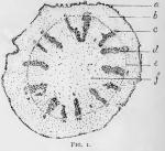 The cross-section of the rhizome or of its branches, when stained by aid of phloroglucin and hydrochloric acid to reveal distinctly the wood wedges, shows that the latter are rather short, irregular in size and placed at unequal distances apart around a large central pith. The vasal bundles are usually considerably narrower than the medullary rays which separate them, and the bark is rather thick. These facts are shown in Fig. 1.
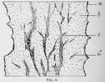 A longitudinal section stained in the same way shows the bundles to be also irregular in their course, and that adjacent bundles frequently send out anastomosing branches, as indicated in Fig. 2.
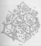
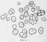 The parenchyma both of the rhizome and roots contain, if the drug is gathered in autumn, as should be the case, a considerable quantity of rather fine-grained starch, as shown in Figs. 3 and 4. The starch grains are more commonly simple and rounded, or somewhat angular, with a central or subcentral not usually conspicuous hilum, and only rarely showing concentric markings. Many of the grains, however, are compound, in twos, threes, or occasionally even in masses composed of several grains, very rarely as many as nine or ten.
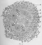 The roots afford an interesting microscopical study and reveal a structure which is quite characteristic. If a section be made a little way back of a root-tip, another near its middle and a third near its base, the primary structure of the central bundle and the secondary changes it undergoes may be easily traced. The primary bundle is usually tetrarch or possesses four xylem rays, but is sometimes triarch or pentarch. Fig. 5 shows a tetrarch bundle from a young portion of a root in which the bundle is but little altered by secondary changes. A wavy zone of cambium has only just been formed between the phloem masses and over the ends of the xylem rays.
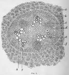
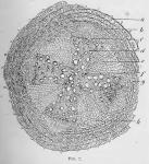 In Fig. 6 the secondary changes have progressed much farther, the whole bundle is much increased in size by growth in the endodermis, in the pericambium and particularly in the cambium zone. The inner ends of the xylem rays have grown by the formation of new ducts until the bases of some of the adjacent rays appear to coalesce. The phloem masses have also increased considerably in size by new growths on their inner face. Finally, in Fig. 7, a section of the old or mature portion of the bundle is shown. In this the bundle is observed to be enormously increased in size, and most conspicuous among the structural changes observed are the formation between each pair of primary xylem rays and back of each phloem mass a large xylem wedge, so that the xylem elements in their arrangement now present the form of a Maltese cross. Alternating with the arms of this cross are four broadly-wedge-shaped medullary rays (also secondary formations), the thin inner end of each wedge resting upon one of the original xylem rays, as shown at f in the figure.
In this species it will be seen that the number of secondary xylem wedges and of medullary rays corresponds to the number of xylem rays and of phloem masses in the primary radial bundle.
The root thus affords us the best characters for the identification of the drug. There are few roots in whifh the most characteristic secondary changes that occur in the roots of dicotyls are traceable with so little difificulty as in this. It therefore affords an especially good example for the young microscopist to study.
It should be observed also that the number of rays is not always constant in the same root. It may, for example, be triarch at the apex and tetrarch near its base, or it may be tetrarch near its apex and pentarch toward its base. In this respect, however, the roots of cimicifuga are not exceptional, many other dicotyls as well as many monocotyls showing similar variations in the number of rays.
The American Journal of Pharmacy, Vol. 67, 1895, was edited by Henry Trimble.
