Some further observations on the structure of Sanguinaria canadensis.
In The Pharmacist for July, 1885, the author published an article on this plant, in which he described the secretion cells and laticiferous tissue of the rhizome.
The following language was used:
"The laticiferous tissue is an interesting subject of study from the morphological standpoint, as in the rhizome it shows every gradation of development from simple, isolated resin- or secretion-cells, through those that are clustered in rows of two or three and those that form an irregular and long chain, but still have a distinct cellular character, to those which form distinct tubes."
A remark of De Bary in which he states that Sanguinaria contains no proper laticiferous tissue, but only secretion cells, led to this re-investigation of the subject. Fresh materials were procured and a large number of sections, longitudinal and transverse, of the rhizome were made and carefully studied with the view of ascertaining with certainty whether or not milk-tissue, in the proper sense of the term, really does exist in the rhizome.
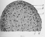
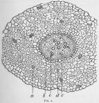
The red or orange-colored secretion is without doubt chiefly contained in distinct cells, which are either isolated or connected into irregular chains and distributed among the parenchymatous tissues of the middle bark and large pith. But in the inner portion of the middle bark, and in the inner bark, occur chains of cells which are longer, more regular and contain a yellow rather than an orange-red secretion. The cells composing the chains are also much narrower and more elongated than are the ordinary secretion cells. Among these rows it is impossible in most instances to demonstrate any communication between the cells. The transverse partitions between the cells are in fact imperforate. In a few instances, however, particularly in the inner layer of the bark, there is demonstrable connection between the secretion cells of the chains, which thus form a true laticiferous tissue, essentially like that occurring in many other of the Papaveraceae, though of course much less complex in its development. It is seldom the case that these milk-tubes are more than a dozen cells long, and they are seldom branching. In fact we find in this plant the form of laticiferous tissue called "complex," or " reticulate," only in the most rudimentary stages of its development. It plays a very subordinate part in holding the secretions of the plant; but still, to the morphologist it is highly significant, as showing the relationship existing between secretion cells and complex laticiferous tissue.
That the secretion cells contain resins beside the alkaloidal principles present in the drug, is clearly evidenced by tests. Moreover, it seems probable that the salts of sanguinarine are more abundant in the large orange-red secretion cells of the pith and outer portion of the middle bark, while those of the closely related alkaloid, chelerythrine, are more abundant in the smaller yellow cells and laticiferous tubes of the inner bark and inner part of the middle bark.


 Sections treated for twenty-four hours or more with strong glycerin showed deposits in the secretion cells of stellate masses of yellowish- brown crystals, with a decided diminution of the inten- sity of color in the liquid contents of the cells. The crystals polarize beautifully, but for lack of time their chemical nature has not been investigated.
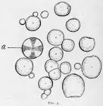
A number of drawings were made in the course of the study, illustrating further the structure of the rhizome and root. These drawings, together with one from the author's Laboratory Exercises, giving a view of the plant and illustrating its floral structure, are reproduced herewith.
The American Journal of Pharmacy, Vol. 67, 1895, was edited by Henry Trimble.


