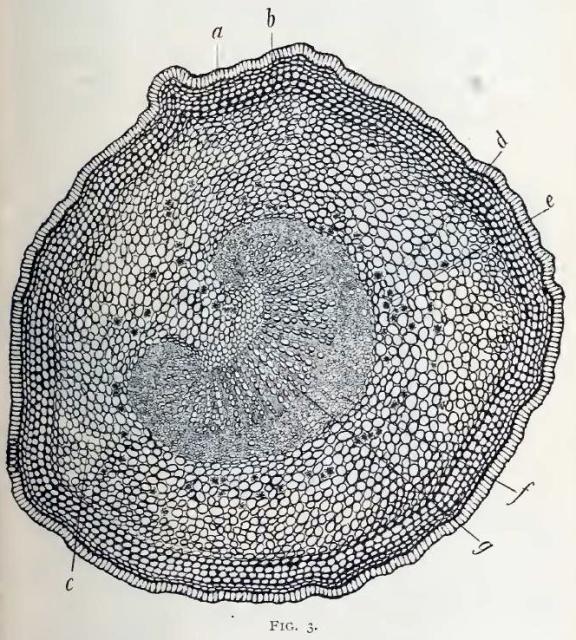
Eriodictyon. Fig. 3.—Transverse section of the petiole
magnified 30 diameters.
a, the epidermis of the petiole, composed of a single layer of cells which are thickened and slightly cutinized upon their exterior surface, and presenting a fringed appearance;
b, several layers of collenchyma or thick angled cells underlying the epidermis;
c, intercellular spaces in the parenchyma;
d, parenchyma tissue surrounding the vasal bundle;
e, crystals of calcium oxalate in the parenchyma cells closely encircling the vasal bundle;
f, phloem portion of the vasal bundle facing the lower surface of the leaf;
g, xylem portion of vasal bundle showing the radial arrangements of ducts.
This image is from Eriodictyon Glutinosum in the November issue of the American Journal of Pharmacy, 1895.


