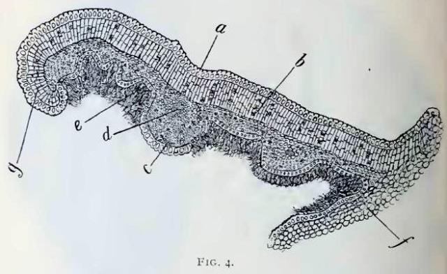
Eriodictyon. Fig. 4.—Transverse section of a portion of lamina adjoining midrib
magnification, 50 diameters;
a, epidermal cells arranged in a single layer, cells very thickwalled;
b, layers of palisade parenchyma composed of several rows of cells and containing crystals of calcium oxalate;
c, spongy parenchyma adjoining lower epidermis of leaf;
d, transverse section through lateral vein, showing small vasal bundle;
e, long, matted hairs upon lower surface;
f, portion of adjoining midrib;
g; slightly revolute margin.
This image is from Eriodictyon Glutinosum in the November issue of the American Journal of Pharmacy, 1895.


