Laboratory Contributions
from the Course Preparatory to Medicine in the University of Pennsylvania.
BY PROF. J. T. ROTHROCK, M. D.
Read at the Pharmaceutical Meeting, January 15, 1884.
Mr. Thomas Ridgway Barker, in examining the ordinary liquorice root (Glycyrrhiza glabra) finds imbedded in parenchyma and in wood, bundles of bast fibres. These bundles have what may be called a bundle sheath in which are found crystals of calcium oxalate, shown to be such by the ordinary tests.
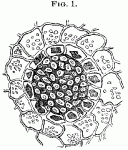 Figure 1 shows the bundle in the parenchyma, seen in cross section.
![]() Figure 2 gives a longitudinal view of the same, divested of its surrounding parenchyma. Figures are magnified about 350 diameters.
Figure 2 gives a longitudinal view of the same, divested of its surrounding parenchyma. Figures are magnified about 350 diameters.
Such crystals and crystal sheaths are not unique. They are found in the Aspidosperma Quebracho, for which see the essay by Dr. Adolph Hansen, reprinted in the "Therapeutic Gazette," October, 1880, p. 292, and are also found in the stem of the anomalous Welwitschia mirabilis, for a figure of which see "De Bary Vergleichende Anatomic," p. 140. There is, however, this difference between the liquorice root and the other plants, i. e. in the former several fibres are included in a single crystal sheath, while in the quebracho and welwitschia there is but a single fibre.
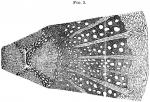 Mr. Jesse G. Shoemaker contributes two diagnostic characters in the stems and roots (say one-fourth of an inch in diameter) of Gelsemium sempervirens, which so far as seen are peculiar in their association, and which hence are of positive value. The first is derived from the medullary rays. These usually widen in a marked manner, going from the centre to the circumference, being sometimes much more than twice as broad exteriorly as interiorly. The second character is the tendency of the pith to be penetrated by several plates of large, thin-walled cells, which divide the pith more or less perfectly into four portions. This latter character, though as far as observed it varies considerably in the relations of the large cells and the ordinary pith cells, is always present and plainly enough marked to serve as a means of diagnosis. Tests upon this point have been made on both fresh and dry specimens received at different times from different places. Figure 3 illustrates these peculiarities, magnified about 400 diameters.
Mr. Charles W. Burr has detected starch in the roots of Coptis trifolia. In Coptis Teeta, Wallich, found in the Mishmi Mountains, eastward of Assam, and recognized by Flückiger and Hanbury as the officinal coptis, starch is known to be present; though in 1873 Mr. E. B. Gross failed to detect it in our American Coptis trifolia. Mr. Burr has repeatedly verified his observation on authentic specimens.
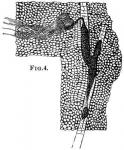 Mr. Wm. C. McFetridge, working upon the Apocynum cannabinum, succeeded in isolating very readily the laticiferous vessels. The illustration shows this quite clearly on longitudinal section (Fig. 4). A transverse section shows the same tissue in a very striking manner, with this difference, that in the latter case the vessels are seen as oval, isolated openings, containing bodies of granular matter inside a very delicate cell wall. There are two special points about these vessels in this species; first the ease with which they may be studied, and second, their relation to the rather anomalous laticiferous vessels in the various cinchona barks.
The American Journal of Pharmacy, Vol. 56, 1884, was edited by John M. Maisch.
