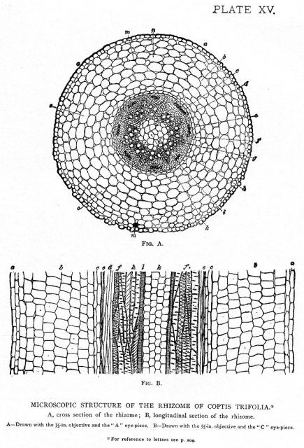
A, cross section of the rhizome; B, longitudinal section of the rhizome.
A—Drawn with the 2/3-in. objective and the "A" eye-piece.
B—Drawn with the 2/3-in. objective and the "C" eye-piece.
LETTER REFERENCES TO PLATE XV AND FIGS. 54, 56, 59, 60, 61, AND 62.—
a, epidermis; b, parenchyma; c, endodermis; d, phloëm; e, liber fibre; f, pitted cells; g, medullary rays; h, wood prosenchyma; i, reticulated cells of parenchyma; k, pith parenchyma; l, spiral vessels; m, resin; n, cells filled with yellow coloring-matter; o, oil-bearing parenchyma; p, palisade tissue; r, cuticle; s, starch or starch-bearing parenchyma; t, root-hairs; v, chlorophyll-bodies; w, stomates.
This image is from Coptis in the Drugs and Medicines of North America.

Henriette's Herbal Homepage
Welcome to the bark side.
Henriette's herbal is one of the oldest and largest herbal medicine sites on the net. It's been online since 1995, and is run by Henriette Kress, a herbalist in Helsinki, Finland.

