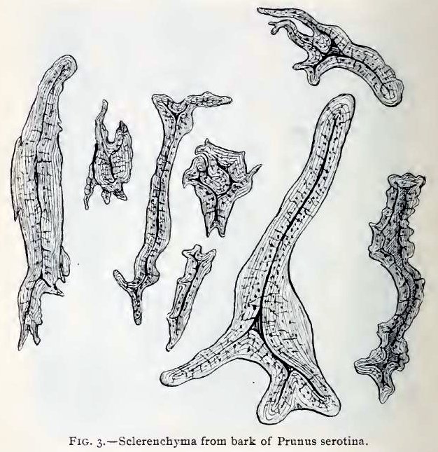
Fig. 3.—Some of the sclerenchymatous elements from the same species, magnified 230 times; the longer of these cells, perhaps, to be regarded as bast fibres, or as transition forms between stone cells and bast fibres.
This image is from Structure of our Cherry Barks in the September issue of the American Journal of Pharmacy, 1895.
