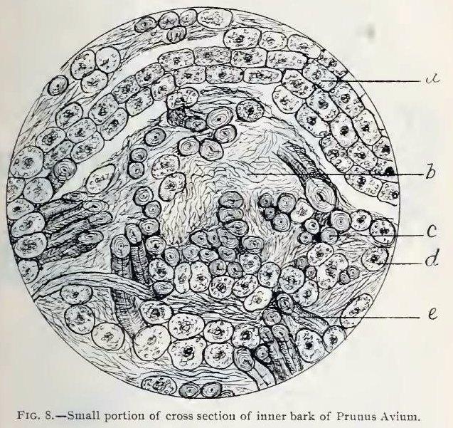
Fig. 8.—Small portion of cross section of inner layer of stem bark of Prunus Avium, magnified about 230 diameters, showing arrangement of bast fibres.
a, portion of medullary ray, well toward the outside of the bast layer;
b, compressed sieve tissues;
c, bast fibre;
d, parenchyma cell;
e, bast fibre, in oblique view.
This image is from Structure of our Cherry Barks in the September issue of the American Journal of Pharmacy, 1895.


