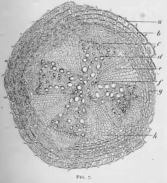
Fig. 7.—Section of central part of a mature root in which the secondary changes have been completed. Magnification, about 60 diameters.
a, parenchyma cell of cortex;
b, cell of endodermis;
c, cambium zone;
d, duct in secondary xylem;
e, broad, wedge-shaped, medullary ray;
f, outer end of one of the original xylem rays at inner end of medullary ray;
h, inter-fascicular cambium. Figs. 5, 6 and 7 are from the author's Laboratory Exercises.
This image is from Structure of Cimicifuga in the American Journal of Pharmacy, 1895.


