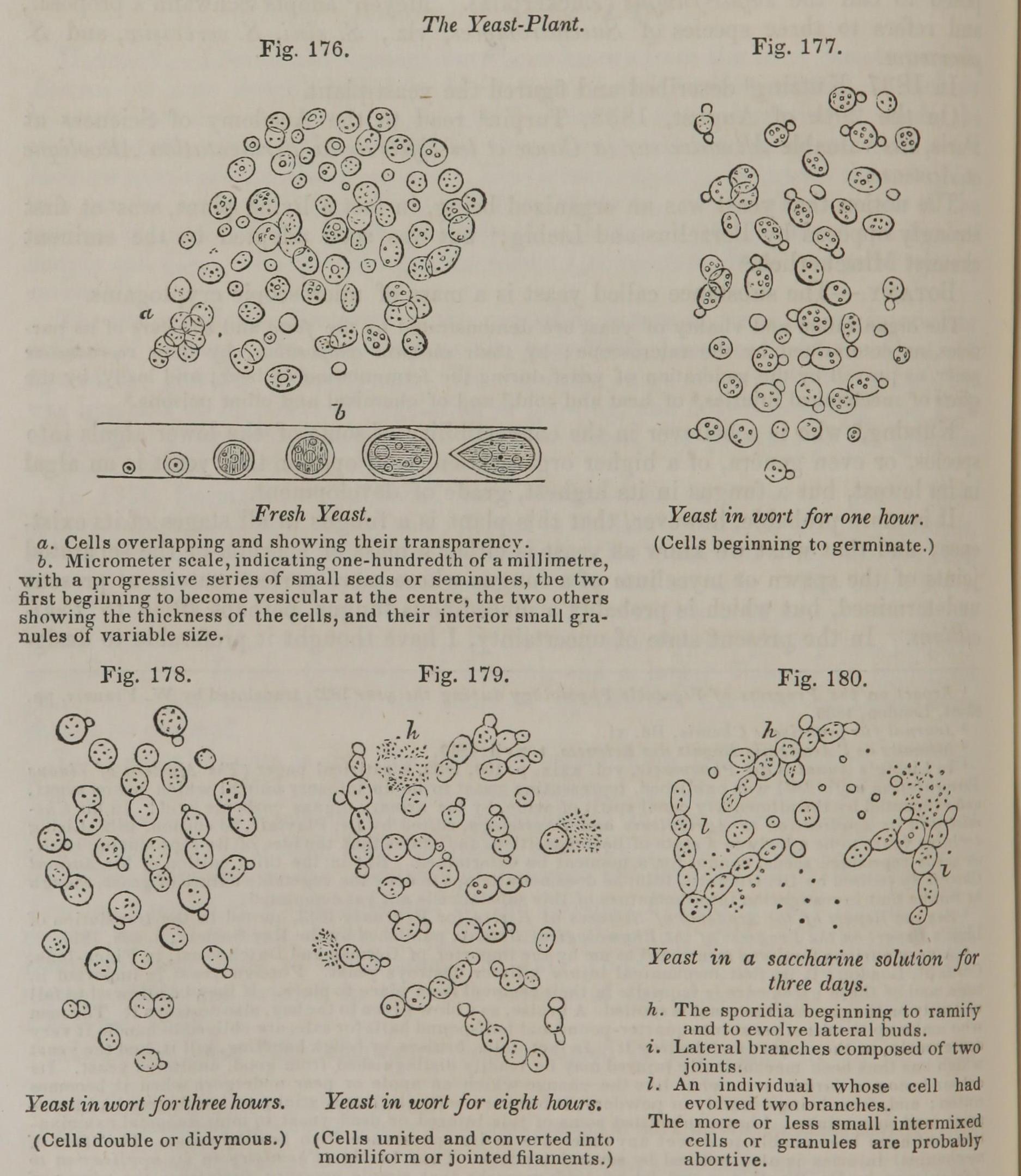
Fig. 176. Fresh Yeast. a. Cells overlapping and showing their transparency. b. Micrometer scale, indicating one-hundredth of a millimetre, with a progressive series of small seeds or seminules, the two first beginning to become vesicular at the centre, the two others showing the thickness of the cells, and their interior small granules of variable size.
Fig. 177. Yeast in wort for one hour. (Cells beginning to germinate.)
Fig. 178. Yeast in wort for three hours. (Cells double or didymous.)
Fig. 179. Yeast in wort for eight hours (Cells united and converted into moniliform or jointed filaments.)
Fig. 180. Yeast in a saccharine solution for three days.
h. The sporidia beginning to ramify and to evolve lateral buds.
i. Lateral branches composed of two joints.
l. An indivitual whose cell had evolved two branches.
The more or less small intermixed cells or granules are probably abortive.
This image is from Yeast in Pereira's Elements of Materia Medica and Therapeutics, 1854.
