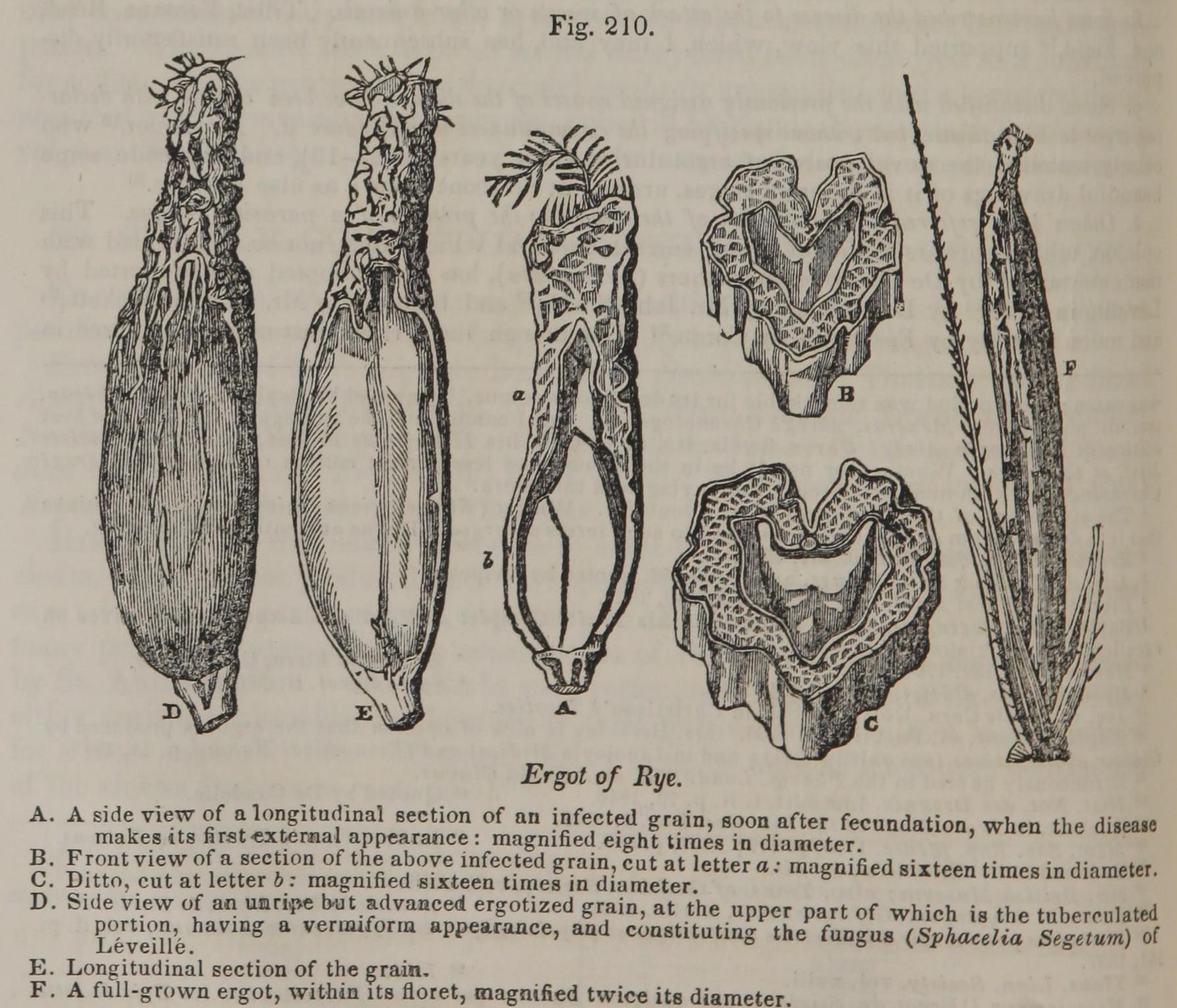
A. A side view of a longitudinal section of au infected grain, soon after fecundation, when the disease makes its first external appearance: magnified eight times in diameter.
B. Front view of a section of the above infected grain, cut at letter a: magnified sixteen times in diameter.
C. Ditto, cut at letter b: magnified sixteen times in diameter.
D. Side view of an unripe but advanced ergotized grain, at the upper part of which is the tuberculated portion, having a vermiform appearance, and constituting the fungus (Sphacelia Segetum) of Léveillé.
E. Longitudinal section of the grain.
F. A full-grown ergot, within its floret, magnified twice its diameter.
This image is from Spurred Rye in Pereira's Elements of Materia Medica and Therapeutics, 1854.
