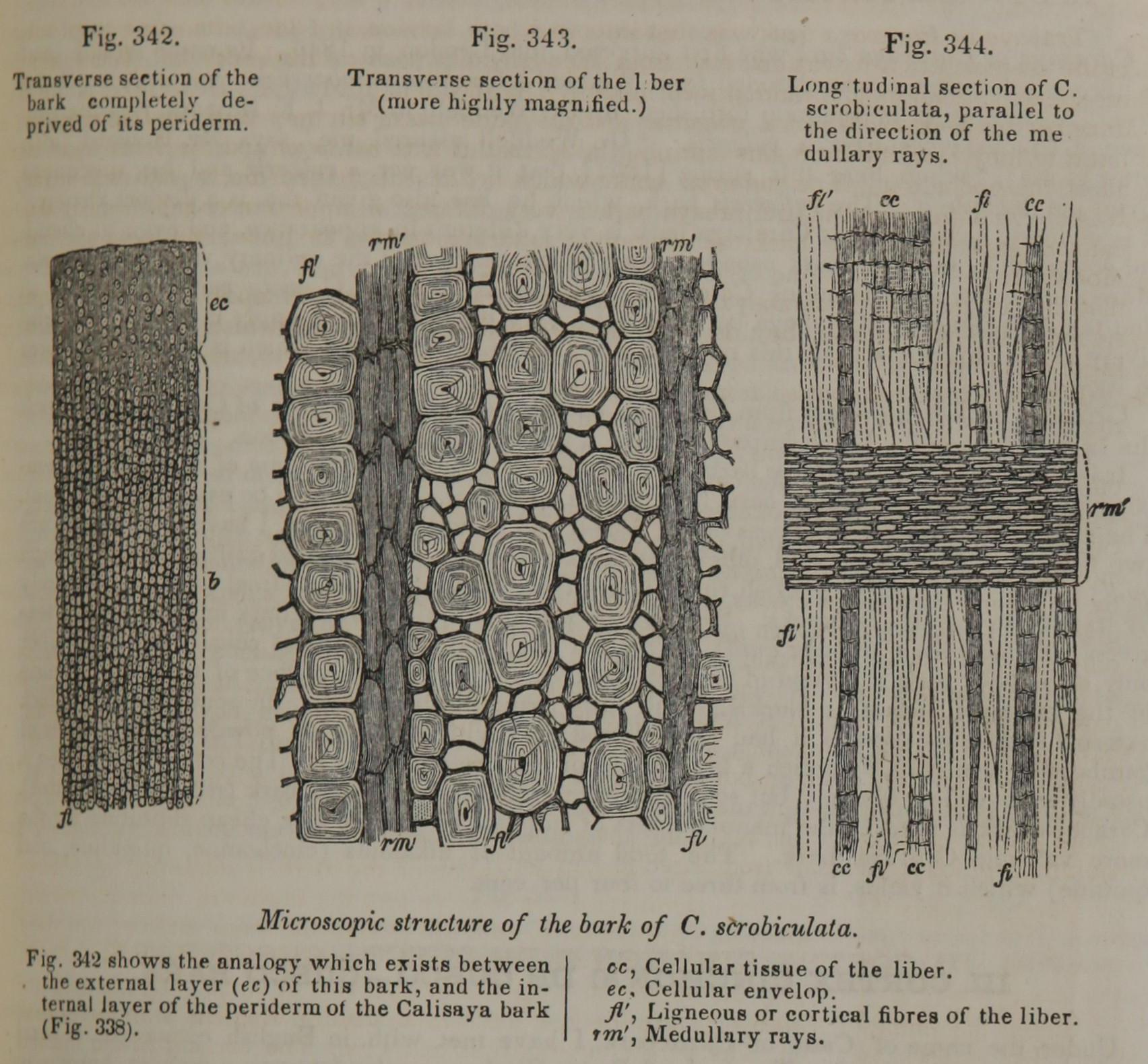
Fig. 342. Transverse section of the bark completely deprived of its periderm.
Fig. 343. Transverse section of the liber (more highly magnified).
Fig. 344. Longitudinal section of C. scrobiculata, parallel to the direction of the medullary rays.
Fig. 342 shows the analogy which exists between the external layer (ec) of this bark, and the internal layer of the periderm of the Calisaya bark (fig. 338).
cc, Cellular tissue of the liber. ec, Cellular envelop. fl', Ligneous or cortical fibres of the liber. rm', Medullary rays.
This image is from _ in Pereira's Elements of Materia Medica and Therapeutics, 1854.
