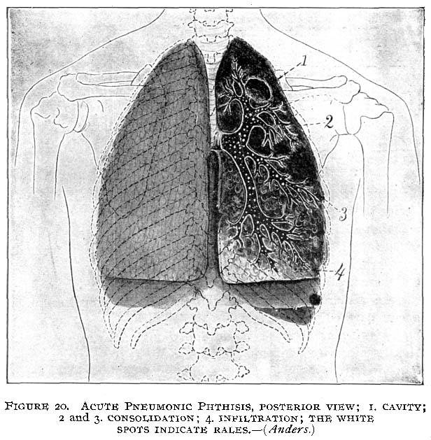
posterior view; 1. Cavity; 2 and 3. Consolidation; 4. Infiltration; The white spots indicate rales. - (Anders.)
This image is from Lobar Pneumonia in Thomas' Eclectic Practice of Medicine.
posterior view; 1. Cavity; 2 and 3. Consolidation; 4. Infiltration; The white spots indicate rales. - (Anders.)
This image is from Lobar Pneumonia in Thomas' Eclectic Practice of Medicine.
Henriette's herbal is one of the oldest and largest herbal medicine sites on the net. It's been online since 1995, and is run by Henriette Kress, a herbalist in Helsinki, Finland.
