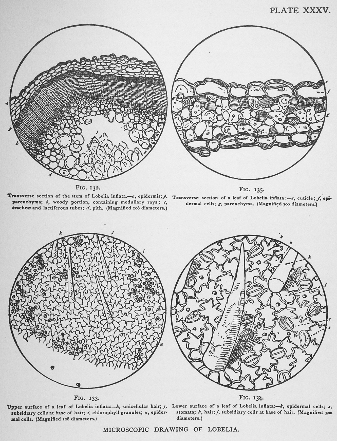
Fig. 132. Transverse section of the stem of Lobelia inflata.—a, epidermis; p, parenchyma; b, woody portion, containing medullary rays; c, tracheae and lactiferous tubes; d, pith. (Magnified 108 diameters.)
Fig. 133. Upper surface of a leaf of Lobelia inflata:—h, unicellular hair; j, subsidiary cells at base of hair; i, chlorophyll granules; n, epidermal cells. (Magnified 108 diameters.)
Fig. 134. Lower surface of a leaf of Lobelia inflata:—k, epidermal cells; s, stomata; h, hair; j, subsidiary cells at base of hair. (Magnified 300 diameters.)
Fig. 135. Transverse section of a leaf of Lobelia inflata:—e, cuticle; f, epidermal cells; g, parenchyma. (Magnified 300 diameters.)
This image is from Lobelia in the Drugs and Medicines of North America.
